
BPC-157 for Joint and Tendon Repair: What the Research Shows
Written bySpartan Research Team
Among all the tissue types studied in the BPC-157 research literature, musculoskeletal applications — specifically tendon and joint repair — represent some of the most consistently documented and mechanistically well-characterized effects. For researchers studying recovery from tendon injuries, ligament damage, and joint tissue degradation, the BPC-157 literature provides a compelling and increasingly detailed picture of how this peptide interacts with connective tissue biology.
This article reviews the published evidence on BPC-157 for joint and tendon repair, with particular focus on the mechanisms that make tendon healing a scientifically logical application, the landmark studies in this area, and what the research literature realistically suggests about timelines and effects.
Why Tendons Are So Difficult to Heal
To understand why BPC-157’s effects on tendon and joint repair are significant, it helps to understand why these tissues heal so poorly under normal conditions.
Tendons and ligaments are characterized by:
- Low cellularity — Relatively few fibroblasts per unit volume compared to muscle or skin
- Poor vascularity — Tendons receive limited blood supply, particularly in the midsubstance region. The Achilles tendon has a notoriously hypovascular zone that corresponds to where most clinical ruptures occur.
- Slow metabolic turnover — Collagen in tendon has a half-life measured in years, not weeks
- Disorganized repair collagen — When tendons do heal, they frequently lay down type III (scar) collagen rather than the type I collagen that gives tendons their tensile strength, resulting in mechanically inferior tissue
These characteristics mean that tendon injuries — from minor partial tears to complete ruptures — heal slowly, often incompletely, and with significant risk of re-injury due to the inferior mechanical properties of repair tissue.
BPC-157’s documented mechanisms directly address the two most critical rate-limiting factors in tendon repair: inadequate blood supply and disorganized collagen deposition.
Mechanisms Relevant to Joint and Tendon Repair
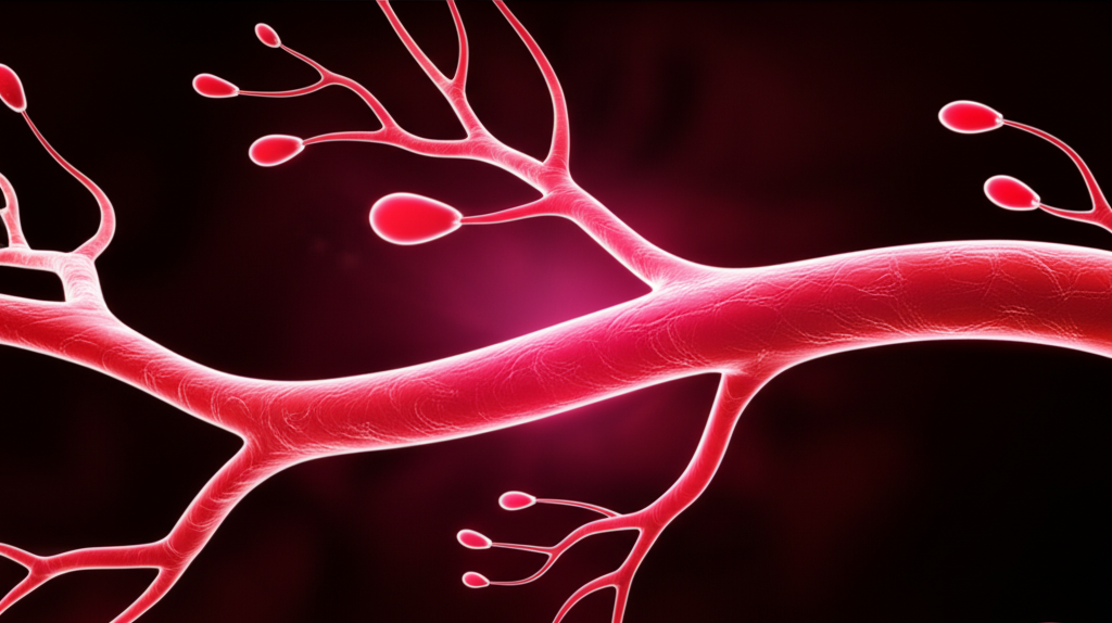
Angiogenesis: Solving the Blood Supply Problem
BPC-157’s most documented mechanism in the tendon repair context is stimulation of angiogenesis — the formation of new blood vessels. Through upregulation of VEGF (Vascular Endothelial Growth Factor) and modulation of downstream angiogenic signaling, BPC-157-treated injury sites in animal models show measurably greater vascularization than untreated controls.
This matters enormously for tendon healing because:
- New vessels deliver oxygen and nutrients to a tissue that is chronically underperfused at baseline
- Improved blood supply delivers the fibroblast precursors and growth factors needed for collagen synthesis
- Greater vascularity in the healing zone is directly correlated with faster maturation and improved mechanical properties of repair tissue
The angiogenic effect of BPC-157 also helps explain why its effects are strongest when administered locally near the injury site — the peptide creates a vascularization gradient centered on the application site, drawing new vessel growth toward the area of maximum tissue demand.
VEGF and NO Pathways: The Vascular-Repair Axis
BPC-157’s pro-healing effects on tendon tissue appear to converge on a VEGF/nitric oxide axis that regulates both vascularization and cellular repair signaling. The interaction works as follows:
BPC-157 → VEGF upregulation → endothelial proliferation → new vessel formation → improved oxygen/nutrient delivery → fibroblast activation → collagen synthesis
Simultaneously, BPC-157’s modulation of the nitric oxide (NO) system normalizes microvascular tone in the healing zone, ensuring that newly formed vessels remain patent and functional rather than regressing or becoming dysfunctional. The NO pathway interaction also contributes to the anti-inflammatory effects observed in tendon healing studies — reducing the prolonged inflammation that can impair collagen organization.
Chang et al. (2011) added an important dimension to this picture by demonstrating that BPC-157 upregulates growth hormone receptor expression in tendon fibroblasts. Growth hormone normally plays a modest direct role in tendon biology, but BPC-157’s receptor upregulation may amplify the angiogenic and collagen-stimulating effects of circulating GH — essentially making tendon fibroblasts more responsive to the endogenous growth signals already present in the healing environment.
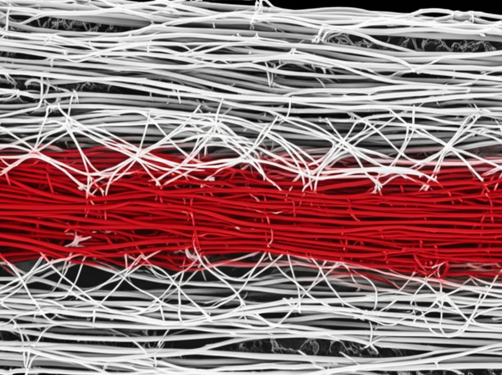
Collagen Synthesis and Organization
Beyond promoting the conditions for healing (vascularity, growth factor availability), BPC-157 appears to directly influence the quality of collagen produced during tendon repair. Published studies report:
- More organized collagen fiber architecture in BPC-157-treated tendons compared to controls
- Evidence of earlier transition from type III (scar) to type I (structural) collagen
- Improved fiber alignment along the tendon’s mechanical axis — the key factor determining tensile strength
This is mechanistically consistent with BPC-157’s activation of the FAK-paxillin pathway, which coordinates fibroblast spatial organization and guides cell-matrix interactions during tissue assembly. When fibroblasts are better organized and better able to sense mechanical cues from the extracellular matrix, they produce more structurally appropriate collagen architecture.
Landmark Studies: The Published Evidence
Achilles Tendon Research
Staresinic et al. (2003) — The Foundational Study
The most-cited study in the BPC-157 tendon literature, published in the Journal of Orthopaedic Research, used a complete Achilles tendon transection model in rats. Animals received BPC-157 (10 μg/kg or 10 ng/kg, intraperitoneally) or vehicle control daily following surgical transection.
Key findings:
- BPC-157-treated tendons showed significantly faster functional recovery (earlier ambulation, less limping)
- Histological analysis revealed better-organized collagen fibers and more cellularity at the repair site
- Biomechanical testing showed improved tensile strength in treated tendons
- Effects were dose-dependent, with both doses showing benefit
- The authors noted particularly strong angiogenic activity at the healing tendon-end interface
This study established the core finding that has been replicated in multiple subsequent experiments: BPC-157 accelerates tendon healing in a manner that produces structurally superior — not just faster — repair tissue.
Chang et al. (2011) — Growth Hormone Receptor Mechanism
Published in Molecules, this study investigated the cellular mechanisms by which BPC-157 exerts its effects in tendon fibroblasts. The critical finding: BPC-157 treatment significantly increased growth hormone receptor (GHR) expression in cultured tendon fibroblasts, particularly under conditions of oxidative stress.
The implications are significant: by upregulating GHR expression, BPC-157 effectively amplifies the tendon fibroblast’s sensitivity to growth hormone signaling. This creates a feedforward loop where BPC-157’s direct effects (improved vascularity, FAK-paxillin activation) are augmented by enhanced responsiveness to endogenous anabolic signals.
The study also documented that BPC-157 protected tendon fibroblasts from hydrogen peroxide-induced apoptosis, suggesting a cell-protective effect relevant to the oxidative stress environment characteristic of acute tendon injury.
MCL (Medial Collateral Ligament) Research
Cerovecki et al. (2010) — Ligament-Specific Data
Published in the Journal of Orthopaedic Research, this study examined BPC-157 specifically in a rat MCL repair model — providing ligament-specific evidence separate from the tendon literature. Complete MCL transection was performed, followed by daily BPC-157 administration.
Results showed:
- Significantly improved functional recovery (hindlimb grip test, gait analysis)
- Superior histological appearance with better collagen organization and higher cellular density
- Increased tensile strength at the repair site in biomechanical testing
- Consistent effect across the dose range tested (10 ng/kg to 10 μg/kg)
The MCL data is particularly valuable because it extends the tendon findings to intra-articular and para-articular ligament structures — suggesting the mechanisms are relevant to joint-associated connective tissue broadly, not just extra-articular tendons.
Application to the Rotator Cuff Context
While dedicated rotator cuff studies using BPC-157 are limited in the published literature, the mechanistic data from Achilles and MCL research applies directly to the rotator cuff’s biological challenges:
- Rotator cuff tendons — particularly the supraspinatus — have a notoriously avascular “critical zone” near the enthesis (tendon-to-bone insertion) where most tears originate and where healing is most impaired
- Post-repair rotator cuff tissue frequently produces disorganized, mechanically inferior scar collagen
- Re-tear rates following rotator cuff repair surgery remain high (up to 25-50% depending on tear size) partly due to inadequate biological healing
BPC-157’s documented ability to enhance vascularization at tendon injury sites and promote organized collagen deposition addresses exactly these failure modes. Research specifically examining BPC-157 in rotator cuff models would represent a high-value next step in the published literature.
Bone-Tendon Interface (Enthesis) Healing
One of the most clinically relevant — and mechanistically challenging — aspects of tendon repair is healing at the enthesis: the tendon-to-bone insertion point. The enthesis is a highly complex, graded tissue that transitions from flexible tendon collagen through fibrocartilage to mineralized bone over just a few millimeters. This complexity makes enthesis healing particularly difficult because the body tends to replace it with disorganized scar tissue rather than recreating the gradient architecture.
Published BPC-157 studies in transection models include data on the enthesis healing zone, and the findings are consistent with the broader tendon data: improved cellular organization, greater vascularity, and better structural integration between the healed tendon end and the bone insertion. The bone healing literature (Sebecic et al., 1999) further supports BPC-157’s ability to enhance osteogenic activity at the repair interface.
Research Timelines: What to Expect
Based on published animal study protocols, the following timeline observations emerge from the BPC-157 tendon/joint healing literature:
Early Phase (Days 1–7)
- Anti-inflammatory effects observed within the first days of administration
- Early angiogenic activity documented (new vessel budding visible in histology)
- Fibroblast migration to the wound site appears accelerated compared to controls
Mid Phase (Days 7–21)
- Significant increase in vascularization at the repair site
- Collagen synthesis actively underway; BPC-157-treated animals show higher type I collagen content at this stage
- Functional improvements in animal models (weight-bearing, ambulation) become measurable
- Growth factor expression (VEGF, FGF, NGF) elevated at the repair site
Late Phase (Days 21–42+)
- Biomechanical advantages become most apparent in testing (tensile strength, stiffness)
- Collagen fiber organization more mature in treated animals versus controls
- Functional recovery scores converge toward normal more rapidly in treated groups
An important caveat: animal models use young, healthy rats with surgically created acute injuries — a context that likely maximizes BPC-157’s observable effect. Human conditions (older age, chronic degeneration, comorbidities, different injury mechanisms) may show different response characteristics. Human timeline data simply does not exist in the current literature.
BPC-157 and the Wolverine Protocol for Joint Research
Many researchers studying joint and tendon repair protocols consider BPC-157 in the context of the Wolverine Protocol — the combination of BPC-157 with TB-500 (Thymosin Beta-4). The rationale is directly relevant to joint healing:
- BPC-157 addresses the local tissue environment — vascularization, fibroblast activation, collagen organization at the injury site
- TB-500 addresses the systemic mobilization problem — driving stem cells and repair cells from circulation into the local healing zone
For complex joint injuries where both local tissue environment and systemic cell recruitment may be limiting factors, the complementary mechanisms of this combination provide a more comprehensive research approach than either compound alone.
Gut Health Considerations in Musculoskeletal Research
An often-overlooked aspect of BPC-157 research in the musculoskeletal context is the peptide’s parallel effects on gut integrity. Gut permeability and systemic inflammation are increasingly recognized as modulators of musculoskeletal healing capacity — chronic low-grade systemic inflammation from gut-derived endotoxins can impair the local healing environment in joints and tendons.
BPC-157’s gut-protective effects (reviewed in our article on Gut Health Peptides) may therefore provide an indirect benefit to joint healing in research models where gut permeability is a variable — an increasingly relevant consideration as the gut-musculoskeletal axis becomes better characterized in the research literature.
Frequently Asked Questions
Does BPC-157 help with joint repair?
Preclinical animal studies suggest BPC-157 may accelerate joint tissue repair through angiogenesis promotion, collagen synthesis stimulation, and growth factor upregulation. Published studies in rat models have shown accelerated healing of tendons and ligaments. Human clinical data remains very limited.
What does the research show about BPC-157 and tendon healing?
Multiple published studies show BPC-157 accelerates tendon healing in animal models. Staresinic et al. (2003) demonstrated faster healing and improved biomechanical strength in transected Achilles tendons. Chang et al. (2011) showed BPC-157 enhances growth hormone receptor expression in tendon fibroblasts, amplifying the regenerative response.
How long does BPC-157 take to work for tendon healing?
In published animal studies, measurable improvements were observed within 2–4 weeks. Anti-inflammatory effects appeared earliest (days 1–7), followed by angiogenic effects (days 7–14), and biomechanical improvements becoming most pronounced at 3–6 weeks. Human timelines are unknown due to limited clinical data.
Can BPC-157 be used for MCL repair research?
Cerovecki et al. (2010) published research specifically on BPC-157 in MCL repair, with treated animals showing significantly improved healing, better tensile strength, and superior collagen organization. This is one of the strongest pieces of evidence for BPC-157’s role in ligament-specific healing.
How does BPC-157 compare to other research compounds for joint repair?
BPC-157 is distinguished by its combination of local angiogenic activity, growth factor upregulation, FAK-paxillin activation, and anti-inflammatory effects — all in a single compound. Other research approaches typically address one mechanism. The Wolverine Protocol combination with TB-500 extends this by adding systemic cell mobilization.
Is BPC-157 safe for research use?
In published animal studies, BPC-157 has shown a favorable safety profile with no organ toxicity, no carcinogenic effects, and no lethal dose established.a> and our dedicated BPC-157 safety profile review.
Key References
- Staresinic M. et al. (2003). “Gastric pentadecapeptide BPC 157 accelerates healing of transected rat Achilles tendon.” Journal of Orthopaedic Research, 21(6), 976-983.
- Chang CH. et al. (2011). “BPC 157 enhances the growth hormone receptor expression in tendon fibroblasts.” Molecules, 16(9), 7954-7963.
- Cerovecki T. et al. (2010). “Pentadecapeptide BPC 157 (PL 14736) improves ligament healing in the rat.” Journal of Orthopaedic Research, 28(8), 1155-1161.
- Sebecic B. et al. (1999). “Osteogenic effect of a gastric pentadecapeptide, BPC-157, on the healing of segmental bone defect in rabbits.” Journal of Orthopaedic Research, 17(4), 480-484.
- Sikiric P. et al. (2018). “Brain-gut Axis and Pentadecapeptide BPC 157.” Current Neuropharmacology, 16(5), 612-641.
- Seiwerth S. et al. (2014). “BPC 157’s effect on healing.” Journal of Physiology and Pharmacology, 65(6), 753-761.
Conclusion
The BPC-157 joint and tendon repair literature is among the most consistent and mechanistically well-characterized bodies of evidence in peptide research. From the foundational Achilles tendon work of Staresinic et al. through the mechanistic detail of Chang et al.’s growth hormone receptor findings and Cerovecki et al.’s ligament data, a coherent picture emerges: BPC-157 directly addresses the core biological deficits that make tendon and ligament healing slow and incomplete.
The convergence of angiogenic promotion, organized collagen synthesis, growth factor upregulation, and anti-inflammatory modulation — all documented in multiple independent studies — provides a mechanistically robust preclinical rationale for continued research into BPC-157’s joint repair applications. The critical next step for the field is well-designed human clinical trials that can determine whether these animal model findings translate to practical tissue repair benefits in human subjects.
For research-grade BPC-157 with third-party purity verification, see Spartan Peptides BPC-157 5mg.
For broader context on BPC-157’s mechanisms and applications,https://spartanpeptides.com/blog/healing-from-within-with-bpc-157/”>Healing from Within with BPC-157, and our overview of BPC-157 for Gut Health.
Research Disclaimer: This article is for research and educational purposes only. BPC-157 is sold exclusively as a research chemical and is not intended for human consumption, diagnosis, treatment, or prevention of any medical condition. All data referenced reflects preclinical animal studies unless otherwise specified. The statements made have not been evaluated by the FDA. Always consult qualified medical and research professionals before conducting any peptide research.
⚠️ Research Use Only — Not for Human Consumption
The peptides discussed in this article are intended for laboratory and research purposes only. They are not intended for human consumption. All information presented is based on published preclinical research and is provided for educational purposes only.
Written by the Spartan Research Team
The Spartan Peptides Research Team consists of scientists, biochemists, and health researchers dedicated to providing accurate, evidence-based information about peptide research. Our content is reviewed for scientific accuracy and updated regularly to reflect the latest findings in peptide science.