
LL-37 Antimicrobial Peptide Research Guide: Cathelicidin Mechanisms

Written bySpartan Research Team
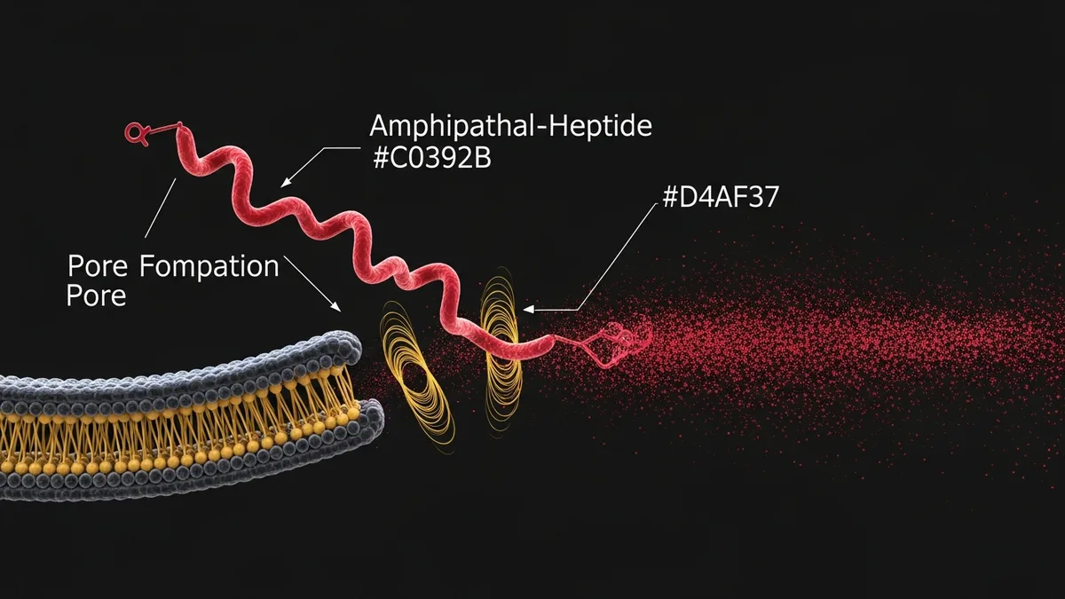
LL-37 is the only known human cathelicidin, a 37-residue cationic amphipathic peptide derived from the C-terminal domain of the hCAP18 (human cationic antimicrobial protein 18 kDa) precursor. Expressed in neutrophils, macrophages, mast cells, natural killer cells, and epithelial surfaces, LL-37 represents a critical component of innate immune defense in research contexts. Its dual role as both a direct antimicrobial agent and an immunomodulatory signaling molecule makes LL-37 a uniquely multifunctional research tool. Researchers use LL-37 to study membrane disruption mechanisms against gram-positive and gram-negative bacteria, biofilm disruption, wound healing modulation, neutrophil extracellular trap (NET) formation, and the immunomodulatory signals through formyl peptide receptor 2 (FPR2) and toll-like receptor pathways.
- Zasloff M (2002) reviewed the mechanism of natural antimicrobial peptides, establishing that cationic helical peptides like LL-37 disrupt bacterial membranes through electrostatic interaction with anionic lipopolysaccharide and phosphatidylglycerol, forming transmembrane pores or causing general membrane dissolution. (PMID 11780977)
- Bowdish DM et al. (2005) documented LL-37 immunomodulatory roles including suppression of LPS-induced TNF-alpha release, chemotaxis induction for neutrophils and monocytes, and induction of IL-1ra, providing a research framework for studying LL-37 as an anti-inflammatory modulatory peptide. (PMID 15653621)
- Research in wound healing models shows LL-37 promotes re-epithelialization by activating EGFR signaling through transactivation, increasing keratinocyte migration and proliferation in scratch-assay and in vivo excisional wound models.
- Biofilm disruption studies show LL-37 penetrates Pseudomonas aeruginosa and Staphylococcus aureus biofilms at concentrations that disrupt the extracellular polysaccharide matrix, reducing biofilm biomass and increasing susceptibility to conventional antibiotics.
- LL-37 expression is regulated by vitamin D, with VDR signaling in keratinocytes and macrophages directly upregulating hCAP18/LL-37 gene transcription, linking nutritional vitamin D status to innate antimicrobial defense in research models.
Membrane Disruption Mechanism
LL-37 adopts an amphipathic alpha-helical secondary structure in membrane-like environments, with hydrophobic residues clustered on one face and cationic residues on the other. This structural feature is critical for membrane disruption activity. In research models, LL-37 follows a carpet model or toroidal pore model of membrane disruption: the peptide accumulates at the bacterial membrane surface through electrostatic attraction to the negatively charged outer leaflet, achieving a threshold surface concentration that destabilizes membrane integrity.
At sub-threshold concentrations, LL-37 disrupts membrane curvature and increases permeability without full lysis. At higher concentrations, rapid membrane dissolution occurs within minutes. The selectivity for bacterial vs. mammalian cell membranes arises from the compositional differences: mammalian membranes are rich in cholesterol and phosphatidylcholine (zwitterionic), while bacterial membranes are rich in anionic phospholipids (phosphatidylglycerol, cardiolipin) that attract the cationic LL-37. Researchers exploit this selectivity in studying LL-37 against gram-positive, gram-negative, and fungal pathogens.
Immunomodulatory Functions Beyond Antimicrobial Activity
Beyond direct membrane disruption, LL-37 functions as an immunomodulatory signaling peptide through multiple receptor interactions. The primary immunomodulatory receptor for LL-37 is formyl peptide receptor 2 (FPR2/ALX), a Gi-coupled GPCR expressed on neutrophils, monocytes, and macrophages. LL-37 activation of FPR2 promotes chemotaxis of immune cells to infection sites and modulates LPS-induced cytokine production, suppressing pro-inflammatory TNF-alpha while enhancing anti-inflammatory IL-1ra output.
LL-37 also interacts with P2X7 receptors and activates intracellular signaling through MAPK and NF-kB pathways, depending on cell type and concentration. At low concentrations in macrophage models, LL-37 can promote anti-inflammatory M2 polarization. At higher concentrations, it activates pro-inflammatory responses. This concentration-dependent immunomodulatory duality is a subject of active research in the antimicrobial peptide literature, with researchers examining the net immunological effect in complex tissue environments.

Wound Healing and Epithelial Research Applications
LL-37 promotes wound repair through mechanisms that extend beyond infection control. In keratinocyte and skin wound models, LL-37 activates epidermal growth factor receptor (EGFR) through transactivation mediated by metalloprotease-dependent release of EGFR ligands. This EGFR activation increases keratinocyte proliferation and directed migration into the wound space, accelerating re-epithelialization in in vitro scratch assays and in vivo excisional wound models.
Angiogenic effects of LL-37 have also been studied in wound healing models. LL-37 activates formyl peptide receptor signaling on endothelial cells, promoting tube formation in Matrigel assays and increasing blood vessel density in subcutaneous tissue models. Researchers studying wound vascularization use LL-37 as a positive control for angiogenic signaling alongside VEGF pathway studies. Spartan does not carry LL-37 itself, but researchers running parallel wound healing protocols often study BPC-157 alongside it for its own tissue repair research profile.
Biofilm Disruption Research
An important research application of LL-37 is in biofilm disruption models. Bacterial biofilms, organized communities encased in an extracellular polysaccharide matrix, are highly resistant to conventional antibiotics. LL-37 penetrates biofilm architecture by disrupting the extracellular matrix components through electrostatic and hydrophobic interactions, while simultaneously disrupting cell membranes of embedded bacteria. Research in Pseudomonas aeruginosa and Staphylococcus aureus models has shown LL-37 reduces biofilm formation at sub-MIC concentrations and disperses established biofilms at higher concentrations.
Combination studies pairing LL-37 with conventional antibiotics (tobramycin, ciprofloxacin) show synergistic activity against biofilm-embedded bacteria in vitro. Researchers hypothesize that LL-37 sensitizes biofilm bacteria to antibiotics by disrupting the protective matrix and increasing membrane permeability, enhancing antibiotic penetration and efficacy.
Research Protocol Considerations
LL-37 is susceptible to proteolysis in serum-containing media, which reduces effective concentration in in vitro studies conducted in the presence of serum. Researchers conducting antibacterial studies typically use low-serum or serum-free buffers for MIC determination and membrane disruption assays. For in vivo wound healing or infection models, the short half-life in tissue is managed by either repeated local injection or formulation in hydrogels or slow-release carriers.
Salt concentration markedly affects LL-37 antimicrobial activity in research assays. High NaCl concentrations (physiological saline) partially inhibit LL-37 membrane disruption by competing with cationic LL-37 for anionic membrane binding sites. This salt sensitivity is an important technical consideration: antibacterial activity measured in standard microbiological broth (e.g., MHB) at physiological salt is lower than in low-salt research buffers. Researchers must report assay salt concentrations to enable comparison across studies.
LL-37 and Innate Immune Signaling Research
LL-37 has become a key research tool for studying the intersection of innate immunity and inflammation. Beyond its direct antimicrobial membrane activity, LL-37 modulates multiple immune cell types including monocytes, dendritic cells, mast cells, and neutrophils. Research using TLR4 reporter systems has demonstrated that LL-37 can both activate and suppress TLR-mediated signaling depending on concentration and cellular context, a duality that makes it particularly interesting in inflammatory disease research models. Researchers investigating the broader innate immunity research space can browse Spartan’s immunity research category for related compounds.
Studies examining LL-37 interaction with lipopolysaccharide (LPS) have shown that LL-37 can neutralize bacterial endotoxin by binding LPS directly, preventing its interaction with TLR4/MD-2 complexes on immune cells. This anti-endotoxin activity is mechanistically distinct from direct membrane disruption and contributes to the in vivo anti-inflammatory effects observed in sepsis model research. Hancock et al. (PMID 11159935) characterized this LPS-neutralization function, establishing a framework for understanding LL-37’s role in limiting excessive inflammatory responses during bacterial infections.
Research in macrophage culture models has identified that LL-37 activates P2X7 purinergic receptors, stimulating inflammasome assembly and IL-1 beta secretion at higher concentrations. At lower, physiologically relevant concentrations, LL-37 has been shown to induce autophagy in macrophages, a cellular process relevant to intracellular pathogen clearance and immune homeostasis research. These bidirectional immune effects underscore the concentration-dependent nature of LL-37 biology and the importance of dose-controlled research design.
Mast cell research has revealed that LL-37 induces degranulation and cytokine release from mast cells, linking cathelicidin expression to allergic and inflammatory signaling cascades. In skin biology research, this property has been investigated in the context of rosacea and atopic dermatitis models, where elevated LL-37 expression is consistently observed. Understanding how LL-37 activates mast cells and keratinocytes informs research into the pathophysiology of inflammatory skin conditions mediated by cathelicidin dysregulation.
LL-37 also serves as a ligand for formyl peptide receptor 2 (FPR2/FPRL1), a G-protein coupled receptor expressed on neutrophils, monocytes, and epithelial cells. Activation of FPR2 by LL-37 promotes neutrophil chemotaxis and transepithelial migration, helping coordinate the innate immune response to tissue injury or infection. Research using FPR2 antagonists has confirmed that this receptor mediates a significant portion of the chemotactic effects of LL-37 in neutrophil recruitment models.
Explore Spartan Peptides’ catalog of research-grade compounds: Browse All Research Peptides at Spartan Peptides
Written by the Spartan Research Team
Our team of peptide researchers and biochemists reviews every article for scientific accuracy. Learn more about our team →