
Thymosin Beta-4: Structure, Function and How TB-500 Relates to It

Written bySpartan Research Team
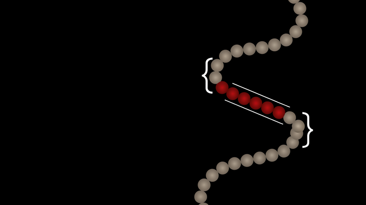
Thymosin beta-4 is a 43-amino-acid peptide found in most mammalian cells and the primary actin-sequestering molecule in eukaryotic cells. TB-500 is a synthetic seven-residue fragment, Ac-LKKTETQ, corresponding to residues 17 to 23, the actin-binding region. The fragment is not identical to the full peptide. Available for laboratory research only.
🔬 KEY RESEARCH FINDINGS
- Not the same molecule: Thymosin beta-4 is the complete 43-residue endogenous peptide. TB-500 is a synthetic seven-residue fragment, Ac-LKKTETQ, matching residues 17 to 23 of the full sequence, characterized directly by Esposito and colleagues (PMID 22962027).
- Actin-sequestering mechanism: Goldstein, Hannappel, and Kleinman describe thymosin beta-4’s core role as binding free G-actin monomers and holding them in reserve, a function that underlies its downstream tissue repair activity (PMID 16099219).
- Accelerated dermal wound closure in rats: Malinda and colleagues reported faster full-thickness wound closure and increased collagen deposition in a rat punch wound model treated with the intact peptide (PMID 10469335).
- Faster corneal healing in mice, with reduced inflammatory cell infiltration: Sosne and colleagues observed accelerated epithelial closure and fewer neutrophils at the injury site in a mouse alkali-burn corneal model (PMID 11950239).
- Not the same peptide as thymosin alpha-1: The two share a thymic origin and a name prefix, but belong to different structural families with different research applications entirely.
What thymosin beta-4 is
Thymosin beta-4 is a small, highly conserved polypeptide made of 43 amino acids, with a molecular weight of roughly 4.9 kDa. It was first isolated from calf thymus tissue in the early 1980s, which is where the “thymosin” name comes from, though later work showed the peptide is not actually thymus-specific. It turns up in nearly every mammalian cell type that’s been checked: platelets, macrophages, epithelial cells, fibroblasts, cardiac tissue, and more.
Structurally, thymosin beta-4 sits in the beta-thymosin family, a group of small proteins that share a common actin-binding fold. It’s intrinsically unstructured in free solution, meaning it doesn’t fold into a fixed rigid shape the way many enzymes do. That flexibility matters mechanistically (more on that below), and it’s also part of why the molecule is easy to confuse with fragments derived from it.
Platelets carry particularly high concentrations of thymosin beta-4, and it’s released during clotting and at wound sites. Concentration in blood plasma and in wound fluid has been measured directly in several of the studies referenced later in this guide. The peptide’s near-universal distribution across cell types is one reason researchers have looked at it across such a wide range of tissue systems, from skin to cornea to heart muscle. This is a general biology and biochemistry entity, not a single-tissue signaling molecule.
One more point worth making early: because thymosin beta-4 (Tβ4) is so widely expressed and evolutionarily conserved across vertebrate species, most of what’s known about its actin-binding role comes from basic cell biology work rather than from a single disease model. That breadth is exactly why the fragment-versus-full-peptide distinction below matters so much. A lot of downstream literature and product copy blurs which molecule is actually being discussed.
Thymosin beta-4 and TB-500 are not the same molecule
This is the point that gets muddled across most peptide research writing, so it’s worth stating plainly: thymosin beta-4 and TB-500 are related, but they are not interchangeable names for the same molecule.
Thymosin beta-4 is the complete, endogenous 43-amino-acid peptide described above. TB-500 is a synthetic fragment, specifically the sequence Ac-LKKTETQ, corresponding to residues 17 through 23 of the full peptide. That’s a seven-residue stretch pulled from the middle of a 43-residue chain. The name “TB-500” itself is not a standardized biochemical designation; it originated as a research and marketing label, and different sources have used it loosely, sometimes to mean the synthetic fragment and sometimes, incorrectly, as shorthand for the full peptide.
Why does this matter beyond terminology? The seven-residue fragment corresponds to the core of the actin-binding domain of thymosin beta-4. Structural and biochemical work on the beta-thymosin fold (Dominguez, 2007; Xue et al., 2007) has mapped how the full peptide wraps around a G-actin monomer using contacts distributed across a larger stretch of the sequence than just residues 17 to 23. A 2012 analytical chemistry paper synthesized and characterized this exact fragment, describing the N-terminal acetylated 17-23 sequence identified in commercial TB-500 material (Esposito et al., 2012). That paper is a useful anchor precisely because it approaches the fragment as a distinct chemical entity worth characterizing on its own, not as a stand-in for the full molecule.
The practical upshot: the fragment retains some actin interaction capacity because it includes part of the binding interface, but it lacks the full three-dimensional context, additional binding contacts, and structural flexibility that the intact 43-residue peptide brings to the interaction. Full regulatory activity attributed to thymosin beta-4 in cell biology, including some of the interactions and signaling roles reported in the tissue studies discussed later, was demonstrated using the intact peptide, not the isolated fragment. Extrapolating full-peptide findings onto the fragment, or vice versa, is not a scientifically sound assumption without direct testing.
| Thymosin beta-4 | TB-500 | |
|---|---|---|
| Length | 43 amino acids | 7 amino acids |
| Sequence | Full endogenous peptide | Ac-LKKTETQ |
| Position | Complete molecule | Residues 17 to 23 |
| Actin-binding domain | Present | Present |
| Full regulatory activity | Yes | Partial |
| Origin | Endogenous, found in most cells | Synthetic fragment |
Spartan Peptides stocks TB-500 research peptide vials alongside reference material and literature notes so researchers can keep the fragment and the full endogenous peptide straight when designing an experiment. Getting this distinction right at the design stage avoids a lot of downstream confusion when comparing results against the published thymosin beta-4 literature.

The actin-sequestering mechanism
The core biochemical role of thymosin beta-4 is binding monomeric actin, also called G-actin, and holding it in a sequestered pool that isn’t available for polymerization into filaments. Goldstein, Hannappel, and Kleinman’s 2005 review in Trends in Molecular Medicine describes this as the peptide’s primary molecular function, with tissue repair activity emerging as a downstream consequence rather than a separate mechanism entirely.
Here’s the short version of how it works. Actin exists in cells in two forms: free monomers (G-actin) and assembled filaments (F-actin). Cells constantly build and break down these filaments to change shape, migrate, and divide. Thymosin beta-4 binds G-actin in a 1:1 complex, holding a portion of the cellular actin monomer pool in reserve rather than letting it immediately polymerize.
Why keep a reserve pool at all? Because rapid, controlled filament assembly and disassembly is what lets a cell change shape and move. A large pool of instantly available monomers, buffered by a sequestering protein like thymosin beta-4, means a cell can respond fast when a filament-nucleating signal fires, without waiting on new actin synthesis. Bubb’s 2003 review in Vitamins and Hormones works through this buffering mechanism in more detail, including how thymosin beta-4’s binding affinity for actin compares with other sequestering proteins in the cell.
This actin regulation connects directly to cell migration. Fibroblasts, keratinocytes, endothelial cells, and immune cells all rely on actin dynamics to move toward a wound site or a chemical gradient. A peptide that modulates the available pool of polymerization-ready actin has an obvious lever on how fast and how far those cells can migrate in a given tissue model. That’s the mechanistic thread connecting an actin-biochemistry finding to the wound and tissue repair research covered in the next section.

It’s worth being precise here: sequestering activity has been characterized using the intact 43-residue peptide. The actin-binding domain within thymosin beta-4 spans roughly residues 4 to 30 depending on which structural model you’re reading, which is part of why the isolated 17-23 fragment (TB-500) retains partial but not complete actin-interaction behavior relative to the full molecule.
Structural modeling work by Xue, Aguda, and Robinson (2007), published alongside Dominguez’s fold review in the same Annals of the New York Academy of Sciences volume, proposed atomic-level models of how beta-thymosins wrap around the actin monomer surface. Their models show multiple contact points distributed across the peptide chain rather than a single short binding motif. That distributed-contact picture is the structural reason a seven-residue slice of the sequence behaves differently from the intact peptide in binding assays. It isn’t that the fragment does nothing; it’s that it’s missing several of the contact points the full chain uses.
What the tissue repair research shows
Two studies anchor most of what’s cited about thymosin beta-4 and tissue repair, and both used the full peptide in animal tissue models, not the synthetic fragment.

Malinda and colleagues (1999) published a rat dermal wound study in the Journal of Investigative Dermatology. Full-thickness skin punch wounds were created on rats, and thymosin beta-4 was applied topically to the wound site. The treated wounds showed accelerated closure compared to untreated controls, along with increased collagen deposition and faster re-epithelialization in the wound bed. The researchers also reported increased recruitment of inflammatory cells and fibroblasts to the site early in the healing timeline, consistent with the actin-driven cell migration mechanism described above. This is a rat skin model. The findings describe wound closure kinetics in that specific animal tissue system, not a claim about any other species or context.
Sosne and colleagues (2002) ran a different model entirely: alkali-induced corneal injury in mice, published in Experimental Eye Research. Alkali burns to the cornea are a standard experimental injury model because they produce a reproducible wound with a strong inflammatory component. In this mouse corneal model, thymosin beta-4 treatment was associated with faster corneal epithelial wound closure and a measurable reduction in the number of infiltrating inflammatory cells (specifically neutrophils) at the injury site, compared to vehicle-treated control eyes. The paper frames this as evidence that thymosin beta-4 activity extends beyond epithelial cell migration alone and includes some modulation of the local inflammatory response, at least in this ocular tissue model.
Two things stand out reading these side by side. First, the tissue systems are completely different: skin versus cornea, rat versus mouse, mechanical punch injury versus chemical alkali burn. The fact that a comparable direction of effect (faster closure, altered inflammatory cell presence) shows up in both supports the idea that the mechanism is a general one tied to actin and cell migration biology, not something specific to one tissue type.
Second, and this is the point that’s easy to lose in summary write-ups: both studies used topical or local application of the intact peptide in a defined animal wound model. Neither paper is describing a human clinical outcome, a systemic effect, or an oral or injectable regimen in a person. Researchers designing follow-up experiments have generally kept close to this framework: local or topical application, a defined animal tissue injury model, and endpoints like wound area, epithelialization rate, and inflammatory cell counts rather than subjective symptom measures.
The Goldstein 2005 review referenced above ties these two specific studies together with the broader actin-sequestering mechanism, which is why it’s cited as the connective reference across sections of this guide rather than as a standalone primary-data source.
Other research directions
Beyond the two anchor tissue models above, thymosin beta-4 shows up across a handful of other preclinical research areas worth flagging, even where the evidence base is thinner or the mechanism less directly tied to actin sequestering.
Corneal research extends past the single alkali-burn study cited above. Sosne and collaborators, along with other groups, have investigated thymosin beta-4 in additional corneal injury and dry-eye models in animals, generally reporting a similar pattern: faster epithelial closure and reduced local inflammatory cell infiltration. The 2012 review by Goldstein, Hannappel, Sosne, and Kleinman in Expert Opinion on Biological Therapy summarizes this corneal work alongside cardiac and dermal findings, describing thymosin beta-4 as what the authors term a multi-functional regenerative peptide based on the range of tissue systems where the actin-sequestering mechanism has produced comparable outcomes.
Cardiac tissue research is a separate thread. Preclinical cardiac injury models (largely rodent) have examined thymosin beta-4’s role in cell survival signaling and vascular cell migration following induced cardiac tissue injury. This line of research draws on the same underlying actin biology but applies it to cardiomyocyte and endothelial progenitor cell behavior rather than epithelial wound closure. It’s a mechanistically distinct question from skin or cornea repair, even though the same peptide and the same core actin-sequestering property are under investigation.
Anti-inflammatory investigation is the third direction. Both the dermal and corneal studies referenced earlier reported changes in inflammatory cell presence at the injury site alongside faster tissue closure, which raised the question of whether thymosin beta-4 has a direct anti-inflammatory signaling role, separate from its effect on cell migration. Crockford and colleagues’ 2010 review in the Annals of the New York Academy of Sciences covers this angle in detail, walking through structural, functional, and biological property data supporting a range of investigated applications, while noting that the relative contribution of direct anti-inflammatory signaling versus faster tissue closure reducing inflammatory cell dwell time is still an open mechanistic question in the literature.
A fourth, smaller thread worth naming: actin oligomer biology more broadly. Work by Qu and colleagues (2015) on distinct actin oligomer states and how different actin-nucleating and actin-binding proteins modulate them provides useful background for anyone trying to place thymosin beta-4’s sequestering role within the wider set of proteins that regulate the actin cytoskeleton. It isn’t the only sequestering protein in the cell, and its relative contribution likely varies by tissue type and by what else is happening at the injury site.
None of these directions currently support a claim about outcomes in human subjects. They represent active areas of animal and cell-culture research building on the actin-sequestering mechanism described earlier in this guide. Because thymosin beta-4’s actin-driven repair mechanism and BPC-157’s cytoprotective and angiogenic mechanisms are frequently investigated in overlapping tissue repair models, researchers comparing the two often look at combined BPC-157 and TB-500 research vials to study both mechanisms side by side within the same experimental design.
Thymosin beta-4 and thymosin alpha-1 are different compounds
A second, smaller point of confusion worth clearing up: thymosin beta-4 and thymosin alpha-1 are not the same peptide, related family name aside.
Thymosin alpha-1 is a 28-amino-acid peptide, also originally isolated from thymus tissue, but it belongs to a different structural family (the alpha-thymosins) with a completely different mechanism. Thymosin alpha-1 research has focused mainly on immune modulation, T-cell activity, and cytokine signaling pathways. Thymosin beta-4, as covered throughout this guide, is an actin-sequestering peptide whose research base centers on cell migration and tissue repair models. They share a name prefix and a thymic origin, and not much else mechanistically.
The “beta” and “alpha” naming reflects two distinct thymosin subfamilies discovered around the same period, not two versions of one compound. Confusing them in a literature search is an easy mistake since both terms return overlapping thymus-biology results, but the receptor interactions, structural fold, and downstream research applications diverge almost completely once you look past the shared name origin.
For a full side-by-side breakdown of how thymosin beta-4 and thymosin alpha-1 differ, including mechanism, structure, and the separate research literatures each compound has generated, see our dedicated comparison guide. That resource goes deeper into the alpha-1 immune-signaling side than this guide does, since the focus here stays on beta-4’s actin biology and its relationship to the TB-500 fragment.
Handling and storage
Thymosin beta-4 and related fragment material ship lyophilized (freeze-dried) and should stay that way until immediately before use in an experiment. Lyophilized peptide is comparatively stable at low temperature: standard guidance is minus 20 degrees Celsius for long-term storage, protected from light and moisture, in the original sealed vial.
Once reconstituted in solution, stability drops considerably. Reconstituted peptide solution should be kept refrigerated at 2 to 8 degrees Celsius and used within the timeframe indicated by the supplier’s documentation, generally a matter of weeks rather than months. Repeated freeze-thaw cycles degrade peptide integrity and should be avoided; aliquoting into single-use volumes before freezing is standard laboratory practice for exactly this reason.
Reconstitution itself is a concentration calculation: the volume of bacteriostatic water or another appropriate diluent needed to add to a given mass of lyophilized peptide to reach a target concentration in solution, expressed in mg/mL. Getting this math wrong is one of the more common sources of inconsistent results across labs running comparable experiments. For the full walkthrough, including worked concentration examples and a calculator, see our guide on reconstituting lyophilised research peptides.
Frequently asked questions
What is thymosin beta-4?
Thymosin beta-4 is a 43-amino-acid peptide found in most mammalian cell types. It functions primarily as the major actin-sequestering protein in eukaryotic cells, meaning it binds and holds free actin monomers in reserve, which influences cell shape change and migration.
Is TB-500 the same as thymosin beta-4?
No. Thymosin beta-4 is the complete 43-amino-acid peptide. TB-500 refers to a synthetic seven-amino-acid fragment, Ac-LKKTETQ, corresponding to residues 17 to 23 of the full peptide. The fragment includes part of the actin-binding region but does not replicate the complete structure or full regulatory activity of the intact molecule.
What does actin sequestering mean?
Actin sequestering describes a protein binding free actin monomers (G-actin) and preventing them from immediately assembling into filaments (F-actin). This keeps a buffered pool of actin ready for rapid deployment when a cell needs to change shape or migrate, which is central to how thymosin beta-4 has been studied in cell migration and tissue repair contexts.
What is the difference between thymosin beta-4 and thymosin alpha-1?
They are structurally and functionally distinct peptides that happen to share a thymic origin and a name prefix. Thymosin alpha-1 is a 28-amino-acid peptide studied mainly for immune modulation and T-cell signaling. Thymosin beta-4 is a 43-amino-acid actin-sequestering peptide studied mainly in tissue repair and cell migration models.
What tissue models has thymosin beta-4 been studied in?
Published preclinical research includes a rat full-thickness dermal wound model (Malinda et al., 1999), a mouse alkali-induced corneal injury model (Sosne et al., 2002), and additional investigation in cardiac tissue and other corneal injury contexts summarized in later review papers. All are animal tissue or cell-culture studies.
How should thymosin beta-4 be stored for research?
Lyophilized peptide should be stored at minus 20 degrees Celsius, protected from light and moisture, until reconstitution. After reconstitution, keep the solution refrigerated at 2 to 8 degrees Celsius, use it within the supplier’s stated window, and avoid repeated freeze-thaw cycles by aliquoting into single-use portions.
Is thymosin beta-4 approved for any medical use?
No. Thymosin beta-4 and TB-500 fragment material are not approved by the FDA or any comparable regulatory body for use in humans or animals outside of a research setting. All material sold by Spartan Peptides is intended strictly for laboratory research use.
References
- Goldstein AL, Hannappel E, Kleinman HK. Thymosin beta4: actin-sequestering protein moonlights to repair injured tissues. Trends Mol Med. 2005 Sep;11(9):421-9. PMID: 16099219
- Malinda KM, Sidhu GS, Mani H, Banaudha K, Maheshwari RK, Goldstein AL, Kleinman HK. Thymosin beta4 accelerates wound healing. J Invest Dermatol. 1999 Sep;113(3):364-8. PMID: 10469335
- Sosne G, Szliter EA, Barrett R, Kernacki KA, Kleinman H, Hazlett LD. Thymosin beta 4 promotes corneal wound healing and decreases inflammation in vivo following alkali injury. Exp Eye Res. 2002 Feb;74(2):293-9. PMID: 11950239
- Goldstein AL, Hannappel E, Sosne G, Kleinman HK. Thymosin beta 4: a multi-functional regenerative peptide. Basic properties and clinical applications. Expert Opin Biol Ther. 2012 Jan;12(1):37-51. PMID: 22074294
- Crockford D, Turjman N, Allan C, Angel J. Thymosin beta4: structure, function, and biological properties supporting current and future clinical applications. Ann N Y Acad Sci. 2010 Apr;1194:179-89. PMID: 20536467
- Esposito S, Deventer K, Goeman J, Van der Eycken J, Van Eenoo P. Synthesis and characterization of the N-terminal acetylated 17-23 fragment of thymosin beta 4 identified in TB-500, a product suspected to possess doping potential. Drug Test Anal. 2012 Sep;4(9):733-8. PMID: 22962027
- Bubb MR. Thymosin beta 4 interactions. Vitam Horm. 2003;66:297-316. PMID: 12852258
- Xue B, Aguda AH, Robinson RC. Models of the actin-bound forms of the beta-thymosins. Ann N Y Acad Sci. 2007 Sep;1112:56-66. PMID: 17468228
- Dominguez R. The beta-thymosin/WH2 fold: multifunctionality and structure. Ann N Y Acad Sci. 2007 Sep;1112:86-94. PMID: 17468236
- Qu Z, Silvan U, Jockusch BM, Aebi U, Schoenenberger CA, Mannherz HG. Distinct actin oligomers modulate differently the activity of actin nucleators. FEBS J. 2015 Oct;282(19):3824-40. PMID: 26194975
Research Use Only: Products referenced in this article are sold strictly for research use only, for in vitro laboratory purposes. They are not drugs, dietary products, or cosmetics, and they are not for human consumption or animal consumption, diagnostic use, or therapeutic use of any kind. Nothing in this article should be interpreted as medical advice or as an endorsement of any use outside a qualified research setting.
Written by the Spartan Research Team
Our team of peptide researchers and biochemists reviews every article for scientific accuracy. Learn more about our team →