
Thymulin Research Guide: Thymic Peptide and Immune Modulation

Written bySpartan Research Team
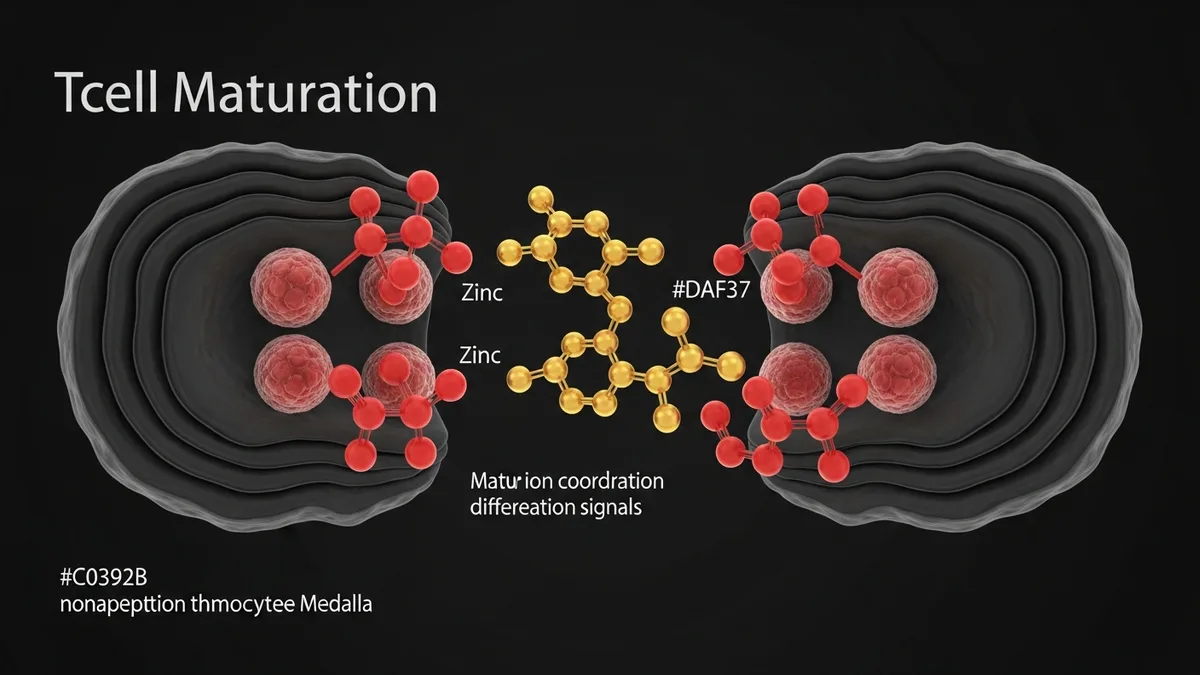
Thymulin is a nonapeptide (Glu-Ala-Lys-Ser-Gln-Gly-Gly-Ser-Asn) produced exclusively by epithelial cells of the thymic cortex and medulla. It is the only thymic peptide known to require zinc for biological activity: thymulin circulates as an inactive apoform and becomes active only when complexed with a zinc ion in a 1:1 ratio. Researchers use thymulin to study T-cell maturation, the immunological thymic clock of aging, and immunomodulatory applications in inflammation and immune deficiency models. Unlike other thymic peptides (thymosin alpha-1, thymopoietin), thymulin’s zinc dependency and exclusive thymic origin make it a specific indicator of thymic function in research contexts.
- Bach JF et al. (1977) first isolated and characterized thymulin (then called thymic serum factor, FTS) from porcine serum, demonstrating that it promotes T-cell differentiation markers in bone marrow cells in vitro. (PMID 340499)
- Dardenne M et al. (1982) demonstrated that thymulin biological activity requires zinc coordination, with zinc-free apoform inactive in T-cell differentiation assays, establishing the zinc-thymulin system as a research tool for studying nutritional immunology. (PMID 7046845)
- Thymulin serum levels decline with age in parallel with thymic involution, with peak levels in early childhood and near-undetectable levels after age 60, making thymulin a biomarker for thymic function in aging research models.
- Zinc deficiency studies show that serum thymulin activity is reduced in zinc-depleted animals, and zinc supplementation restores thymulin bioactivity, linking nutritional zinc status to T-cell immune competence through the thymulin system.
- Anti-inflammatory research in rodent models (experimental arthritis, colitis, neuroinflammation) has documented thymulin-mediated reduction in pro-inflammatory cytokine production (TNF-alpha, IL-1beta, IL-6) and attenuation of disease severity.
Zinc-Dependent Activation Mechanism
Thymulin circulates in blood in two forms: an inactive apoform (zinc-free) and a biologically active zinc-coordinated holoform (Zn-thymulin). The zinc ion forms coordination bonds with specific residues in the nonapeptide structure, inducing a conformational change that enables receptor binding and biological activity. In research assays, thymulin activity is measured by the ability to induce T-lymphocyte markers (TL antigen, glucocorticoid sensitivity, E-rosette formation) on immature T-cell precursors from bone marrow or peripheral blood.
The zinc-thymulin system provides a unique research tool for studying the intersection of nutritional status and immune function. Because thymulin requires zinc for activity, studies of zinc deficiency consistently show reduced effective thymulin signaling even when total thymulin peptide levels are preserved. Conversely, in vitro addition of zinc to serum from zinc-deficient subjects restores thymulin activity, providing a functional assay that differentiates thymulin production defects from zinc-dependent activation defects.
T-Cell Maturation Signaling
Thymulin acts on immature T-cell precursors (thymocytes) and peripheral blood T-cell precursors to promote differentiation markers characteristic of mature T-lymphocytes. In research models, zinc-thymulin treatment induces expression of CD4, CD8, and thymic leukemia (TL) antigen on immature T-cell populations, promotes glucocorticoid sensitivity (a property of mature thymocytes), and enhances E-rosette formation (a functional T-cell competence assay). These effects occur at picomolar to nanomolar concentrations in vitro.
The receptor for thymulin on thymocytes has been described as a cell-surface protein that triggers intracellular signaling leading to cAMP elevation and PKA activation. Downstream signaling promotes gene expression changes consistent with T-cell maturation, including expression of mature T-cell surface antigens and cytokine responsiveness. Researchers studying thymic education and central tolerance mechanisms use thymulin as a tool to modulate thymocyte developmental stage in ex vivo thymic organ culture models.

Thymulin and Aging Research
Thymic involution with age is a well-documented phenomenon in which the thymus progressively replaces functional thymic tissue with adipose and fibrotic tissue, reducing naive T-cell output. Thymulin serum levels parallel this involution, peaking in early childhood and declining progressively to near-zero in elderly subjects. This age-dependent decline in thymulin is studied as both a biomarker of immune aging and a potential research target for immune rejuvenation strategies.
In aged rodent models, thymulin administration has been reported to partially restore T-cell immune competence, increase CD4/CD8 ratios, and improve antibody response to experimental vaccination. Researchers studying thymosin alpha-1 alongside thymulin note complementary mechanisms: thymosin alpha-1 promotes T-cell activation and cytokine production, while thymulin acts earlier in the maturation cascade to support T-cell differentiation from precursors. Spartan does not carry thymulin itself, but researchers studying the thymic peptide family can review the thymosin alpha-1 product page above as the closest available comparison compound, or the Immune Support Research Stack for a combined thymic and immune research protocol.
Anti-Inflammatory Research Applications
Beyond T-cell maturation, thymulin has been studied in inflammation models where it exhibits anti-inflammatory properties independent of its T-cell maturation functions. In rodent experimental arthritis models, thymulin administration reduces joint inflammation, paw edema, and pro-inflammatory cytokine levels (TNF-alpha, IL-1beta). The anti-inflammatory mechanism involves thymulin modulation of macrophage cytokine production and potentially direct interaction with neuroimmune pathways, including evidence for thymulin effects on hypothalamic-pituitary-adrenal axis signaling in neuroendocrine research contexts.
Neuroinflammation models have also been a subject of thymulin research. Published preclinical literature describes thymulin effects on pain perception in nociceptive models, with intrathecal administration reducing inflammatory hyperalgesia. Researchers studying the intersection of immune signaling and pain processing have characterized thymulin as a peripheral and central immunomodulatory peptide with potential relevance to neuroimmune research beyond classical thymic function.
Thymulin Assay Methods and Research Protocols
The standard bioassay for thymulin activity is the rosette inhibition test or the E-rosette induction assay using bone marrow null cells or peripheral blood null cells. These assays measure the ability of test serum or exogenous thymulin to induce T-cell markers on immature precursors, reporting activity as the dilution endpoint at which T-cell differentiation induction is observed.
For in vivo research, thymulin is administered by subcutaneous or intraperitoneal injection in rodent models. The very short half-life of thymulin in circulation (minutes) means that frequent dosing or slow-release formulations are used in chronic administration studies. Researchers comparing thymulin to other immune peptides such as thymosin alpha-1 note the importance of zinc supplementation in thymulin studies to ensure zinc availability for active holoform formation in vivo.
Thymulin in Age-Related Immune Decline Research
Thymulin is among the most directly studied thymic peptides in the context of immunosenescence, the progressive decline in immune function associated with aging. As the thymus involutes through adulthood, circulating thymulin levels decline measurably, and this reduction correlates in research models with decreased naive T-cell output, impaired T-cell proliferative responses, and reduced cytokine production upon antigen challenge. Researchers use thymulin as a biomarker of thymic activity and as an experimental intervention in models designed to study or reverse immune aging.
Studies in aged rodent models have administered exogenous thymulin or zinc supplementation (to support endogenous thymulin activation) and measured immunological endpoints including T-cell subset ratios, cytotoxic lymphocyte activity, and antibody titers following vaccination challenges. Published research from the Mocchegiani group in Italy documented improvements in T-cell function and reduced infection susceptibility in aged mice following thymulin-related interventions, establishing a scientific basis for continued investigation of thymulin in aging research.
Research comparing thymulin levels in young versus aged subjects across animal species consistently finds reductions of 80-90% by midlife, with the decline beginning in late adolescence following peak thymic activity. This dramatic reduction makes thymulin restoration an attractive experimental target in immunosenescence research. Investigators using thymulin as a research intervention typically use bioassays measuring T-lymphocyte rosette formation or flow cytometry-based T-cell marker expression to quantify biological activity of exogenously administered thymulin preparations.
The role of zinc in thymulin biology creates an important intersection with nutritional immunology research. Studies have demonstrated that zinc supplementation in zinc-deficient elderly populations partially restores measurable thymulin bioactivity in serum, consistent with the model that age-related zinc insufficiency contributes to functional thymulin inactivation. Research designs that distinguish between reduced thymulin synthesis (central thymic failure) and reduced thymulin activation (peripheral zinc deficiency) provide important mechanistic clarity for interpreting aging immune data.
Thymulin’s interaction with neuroendocrine systems is another active area of investigation. Research has documented bidirectional communication between the thymic epithelium and the hypothalamic-pituitary axis, with growth hormone and prolactin receptors expressed on thymic epithelial cells. Thymulin in turn influences neuroendocrine signaling, creating feedback loops studied in psychoneuroimmunology research. This bidirectional cross-talk suggests that immune restoration through thymulin research may have implications beyond strict immunological endpoints.
Explore Spartan Peptides’ catalog of research-grade compounds: Explore Spartan Peptides Research Compound Catalog
Written by the Spartan Research Team
Our team of peptide researchers and biochemists reviews every article for scientific accuracy. Learn more about our team →