
PEG-MGF Research Guide: Mechano Growth Factor in Muscle Models
Written bySpartan Research Team
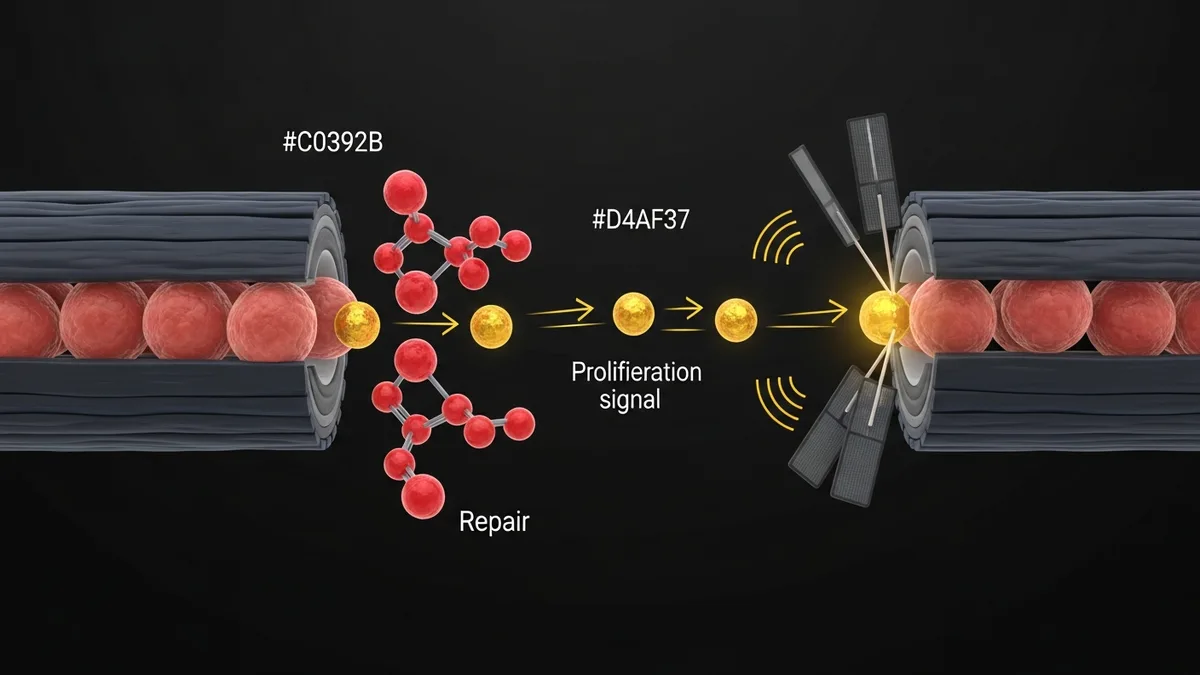
Mechano Growth Factor (MGF) is a splice variant of IGF-1 produced by skeletal muscle in response to mechanical loading or tissue damage. Unlike systemic IGF-1, MGF is expressed locally at sites of muscle stress and functions as an autocrine/paracrine signal that activates muscle satellite cells (muscle stem cells), triggering proliferation and differentiation required for muscle hypertrophy and repair. PEG-MGF is a pegylated form of the MGF E-domain peptide, in which polyethylene glycol (PEG) chains are attached to extend the half-life from minutes to approximately 24 hours, making it practical for preclinical research protocols. Researchers use PEG-MGF to study satellite cell activation, anabolic signaling in muscle injury models, and the distinct role of the IGF-1 Eb (MGF) splice variant vs. systemic IGF-1 Ea.
- Yang SY and Goldspink G (2002) demonstrated that MGF E-domain peptide is functionally distinct from mature IGF-1, activating satellite cell proliferation through a different receptor interaction from the classical IGF-1R. (PMID 12169467)
- Goldspink G (2005) reviewed evidence that MGF operates via an IGF-1-independent signaling mechanism in the early phase of muscle damage response, activating satellite cell entry into the cell cycle before systemic IGF-1 Ea drives later differentiation phases. (PMID 16313558)
- Pegylation of MGF E-domain extends half-life from approximately 5 minutes (native MGF) to approximately 24 hours, enabling single-dose research protocols in animal models that mimic sustained satellite cell activation.
- Preclinical studies in rodent muscle injury models show PEG-MGF increases satellite cell number, myofiber cross-sectional area, and markers of muscle regeneration compared to controls.
- PEG-MGF and IGF-1 LR3 are studied as complementary rather than redundant compounds, with MGF activating satellite cell proliferation and IGF-1 driving subsequent differentiation and protein synthesis.
MGF: The Mechano-Responsive IGF-1 Splice Variant
The IGF-1 gene undergoes alternative splicing to produce multiple isoforms. The class II IGF-1Eb isoform (MGF in rodents, IGF-1Ec in humans) is distinguished by a unique C-terminal E-domain that is not present in the IGF-1 Ea isoform responsible for systemic growth factor activity. When skeletal muscle is mechanically loaded or damaged, local expression of the MGF splice variant increases rapidly, producing the MGF precursor. Proteolytic processing releases the E-domain peptide (the MGF peptide used in research) and mature IGF-1 (identical to systemic IGF-1 Ea in sequence).
The MGF E-domain peptide acts on satellite cells through a receptor interaction that appears independent of the canonical IGF-1 receptor (IGF-1R). Research by Yang and Goldspink (2002) demonstrated that MGF peptide activates satellite cell entry into the cell cycle and promotes nuclear translocation in a manner not blocked by IGF-1R antibodies, suggesting a distinct receptor or co-receptor interaction. This mechanistic distinction is why researchers study PEG-MGF and IGF-1 as complementary tools rather than interchangeable GF analogs.
PEG-MGF vs. IGF-1 LR3 in Muscle Research
Both PEG-MGF and IGF-1 LR3 are studied in muscle anabolic and repair research, but they operate through different phases of the muscle regeneration cascade. PEG-MGF activates the satellite cell proliferation phase, increasing the pool of myogenic progenitors available for repair or hypertrophy. IGF-1 LR3 drives the subsequent differentiation and fusion phase, activating the PI3K/Akt/mTOR protein synthesis pathway in existing and newly formed myofibers.
Researchers designing sequential treatment protocols in muscle damage models have examined PEG-MGF followed by IGF-1 LR3 administration to amplify both phases of the regeneration response. Initial PEG-MGF administration increases satellite cell number (proliferation), and subsequent IGF-1 LR3 administration promotes differentiation and protein anabolism. The temporal separation of these signals aligns with the biological sequence of satellite cell activation, proliferation, differentiation, and fusion observed in in vivo muscle repair.

Satellite Cell Biology in PEG-MGF Research
Satellite cells are quiescent muscle stem cells positioned between the basal lamina and the sarcolemma of mature myofibers. In healthy muscle they are maintained in a G0 quiescent state. Muscle damage or mechanical overload triggers satellite cell activation, characterized by expression of myogenic regulatory factors (MRFs) including MyoD and Myf5, followed by proliferation and differentiation into new myonuclei or repair of damaged fibers.
PEG-MGF research models typically evaluate satellite cell response using immunohistochemistry markers (Pax7, MyoD, Ki67 for proliferation), BrdU incorporation assays, or flow cytometry on dissociated muscle. In rodent models of eccentric exercise-induced injury or cardiotoxin injection, PEG-MGF administration has been reported to increase Pax7-positive satellite cell count and Ki67-positive proliferating cells at 24-72 hours post-treatment. These cellular endpoints are the primary outcome measures in PEG-MGF efficacy studies.
PEG-MGF Research Protocol Design
PEG-MGF is administered by subcutaneous or intramuscular injection in preclinical research. The extended half-life from pegylation allows single-dose or low-frequency dosing in models where native MGF would require continuous infusion. Researchers typically administer PEG-MGF within 24-48 hours of the initiating mechanical or chemical muscle damage event, coinciding with the window of maximal satellite cell activation.
Dose-response studies in rodent muscle injury models have used a range of concentrations, with researchers measuring both satellite cell markers and functional outcomes (grip strength, specific force, myofiber CSA) at timepoints of 7, 14, and 28 days post-injury. For comparison with the BPC-157 and TB-500 recovery stack studied in combined recovery protocol research, PEG-MGF represents a more targeted satellite cell-focused anabolic signal rather than a broad tissue repair agent.
Research Safety Profile
Preclinical safety assessments of PEG-MGF in rodent models show no reported hepatotoxicity, nephrotoxicity, or histological abnormalities in treated animals at research doses. The pegylation modification reduces immunogenicity compared to unmodified peptides by shielding the peptide core from immune recognition. Researchers note that PEG-based modifications are well-characterized in pharmaceutical literature and are considered acceptable excipients in preclinical research design.
PEG-MGF in Tendon and Connective Tissue Research
While PEG-MGF research has focused substantially on skeletal muscle, investigators have also explored its effects in tendon, ligament, and connective tissue models. These tissues contain tenocytes and fibroblasts that express IGF-1 receptors and respond to IGF-1 splice variant signaling. Research in tendon injury models has assessed whether PEG-MGF can modulate collagen synthesis, tenocyte proliferation, and extracellular matrix remodeling following mechanical or chemical-induced tendon damage.
Tenocyte culture experiments have demonstrated that MGF Ec peptide fragments stimulate collagen type I synthesis and tenocyte migration in scratch assay models. These in vitro findings suggest that the E-domain of MGF carries biological activity relevant to connective tissue repair, independent of the IGF-1 receptor binding activity attributed to the mature MGF domain. The pegylation modification in PEG-MGF extends peptide half-life sufficiently to reach connective tissue depots via systemic delivery, making it useful in in vivo injury repair research designs.
Research comparing PEG-MGF to other growth factors in tendon healing models indicates that the mechanically responsive origin of MGF expression is particularly relevant in load-bearing tissue research. Studies using mechanical stretch models in cell culture have shown that MGF expression is upregulated in tenocytes subjected to cyclic tensile loading, paralleling the mechano-responsiveness documented in skeletal muscle. This tissue-level responsiveness makes PEG-MGF a subject of interest in biomechanics and orthopedic biology research.
In bone research, IGF-1 signaling through the IGF-1 receptor is well-established as a regulator of osteoblast activity and bone matrix deposition. PEG-MGF studies in bone cell models have examined whether the splice variant retains osteogenic signaling capacity. Published data from osteoblast culture experiments indicate that MGF Ec peptide exposure increases alkaline phosphatase activity and osteocalcin expression, markers of osteoblastic differentiation, supporting further investigation of PEG-MGF in bone biology research applications.
The systemic bioavailability of PEG-MGF following subcutaneous administration in rodent models has been characterized in pharmacokinetic studies. These studies measure serum concentrations at defined time points using ELISA-based assays specific for the Ec peptide region. Peak concentrations are typically achieved within 15-30 minutes of administration, with the polyethylene glycol modification extending the terminal half-life to approximately 60 minutes compared to less than 5 minutes for unmodified MGF in plasma degradation assays.
Explore Spartan Peptides’ catalog of research-grade compounds: Browse Spartan Peptides Research Compound Catalog
Written by the Spartan Research Team
Our team of peptide researchers and biochemists reviews every article for scientific accuracy. Learn more about our team →